Our Technology
Precision Starts with the Right Tools.
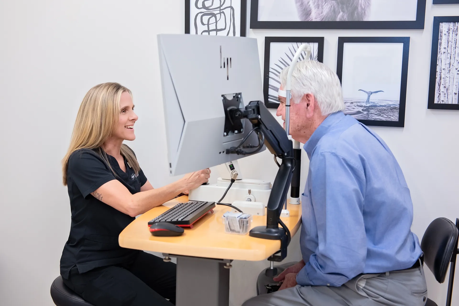
Every surgery begins long before the operating room — with the advanced diagnostic technology that maps your eyes down to the micron.
Schedule Your ConsultationTechnology-Driven Care
Why Technology Matters in Eye Surgery
The difference between a good outcome and a great one often comes down to the precision of your surgeon’s measurements. At Soni Vision Institute, we invest in the most advanced diagnostic and surgical platforms because better data means better decisions — and better decisions mean better vision.
Every measurement, every calculation, every incision is guided by technology designed to deliver the most accurate results possible. From the moment you walk in for your evaluation to the moment your surgery is complete, precision is built into every step of your care.
Our Equipment
Built for Precision. Designed for You.
The diagnostic and surgical platforms we use are among the most advanced available in ophthalmology today.
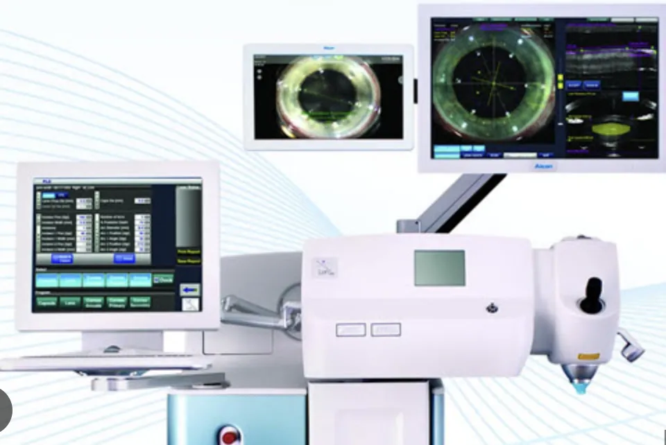
LenSx Femtosecond Laser
by Alcon
The LenSx laser replaces traditional blades with computer-guided femtosecond laser pulses, creating incisions with micron-level accuracy. This technology allows our surgeons to perform the most critical steps of cataract surgery — corneal incisions, capsulotomy, and lens fragmentation — with a level of precision that manual techniques cannot match.
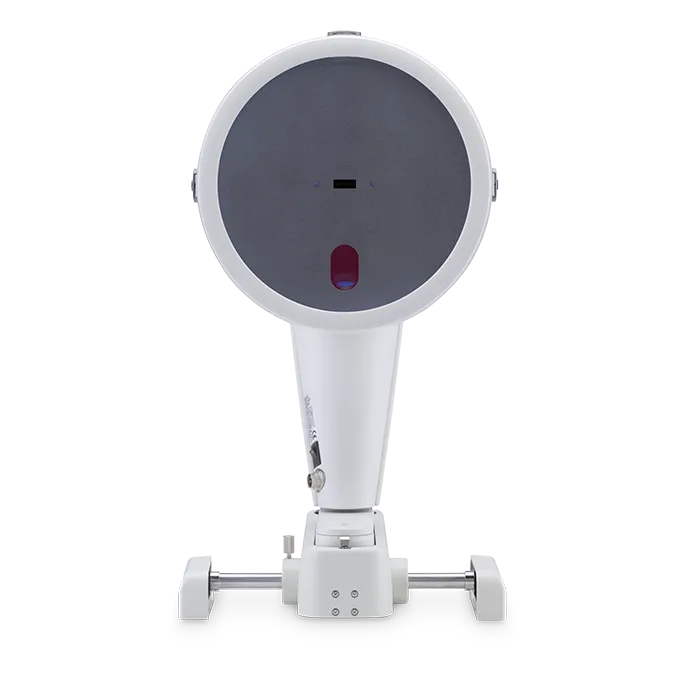
Pentacam HR
by OCULUS
The Pentacam HR uses rotating Scheimpflug camera technology to capture a complete three-dimensional image of the front of your eye in under two seconds. It maps the shape, thickness, and curvature of your cornea from front to back — critical data for selecting the right lens implant and planning refractive procedures.
Corneal Topography
Corneal topography creates a detailed color-coded map of your cornea’s surface curvature — similar to a topographic map of terrain. This data reveals astigmatism patterns, corneal irregularities, and subtle surface conditions that affect surgical planning, lens selection, and contact lens fitting.
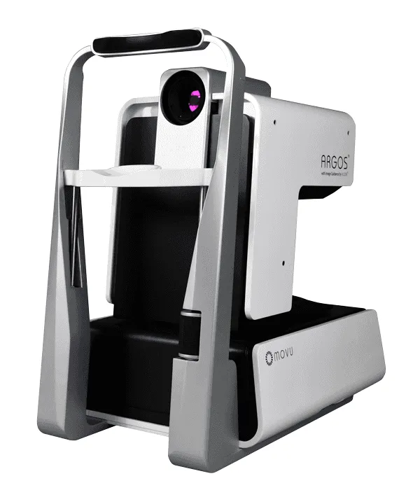
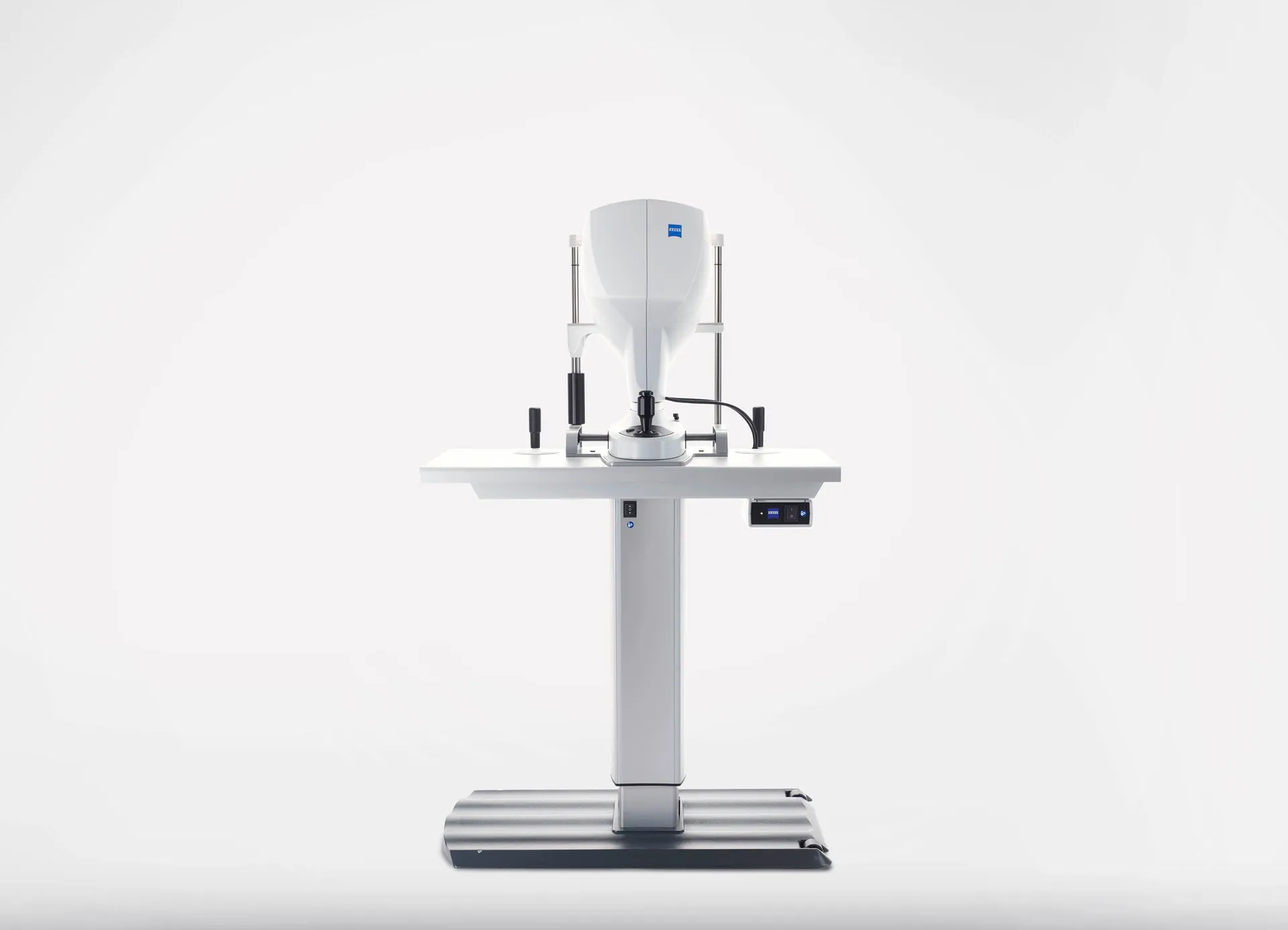
Argos Biometer
by Alcon
The Argos uses swept-source optical coherence tomography (SS-OCT) to measure the precise dimensions of your eye — including axial length, corneal curvature, lens thickness, and anterior chamber depth. These measurements directly determine the power of your intraocular lens implant, making accuracy here essential to your visual outcome.
From Measurement to Surgery — Seamlessly Connected
Measure
Advanced diagnostics capture thousands of data points about your eye’s unique anatomy.
Plan
Your surgeon analyzes the data to select the ideal lens and surgical approach for your goals.
Execute
Computer-guided laser technology delivers the surgical plan with micron-level accuracy.
Experience the Difference Technology Makes
See what’s possible when world-class surgeons meet world-class technology.